Breast Cancer – The Diagnosis Process
Breast Cancer 101
Breast cancer occurs when breast cells begin to grow and divide at an uncontrolled pace.
Normal breast cells grow and divide as needed and then die as they age or suffer damage.
In contrast, breast cancer cells continue to divide, they are resistant to signals that induce cell death, and gain the capacity to invade nearby tissues, eventually forming masses called tumours.

Types of Breast Cancer
I am only going to speak to the five most common types of Breast Cancer here
-
-
- Ductal Carcinoma in Situ (DCIS) refers to abnormal cells found within the milk ducts that are considered non-invasive as the cells have not spread from the ducts to the surrounding breast tissue.
- This is an early form of cancer that in some cases could potentially become invasive and spread to other tissues.
- Lobular Carcinoma in Situ (LCIS) refers to abnormal cells found within the lobules, the milk-producing glands, of the breast.
- While it is uncommon for LCIS to develop into invasive cancer, this condition is linked to an increased risk of developing invasive breast cancer later in life.
-
-
-
- Invasive ductal carcinoma is a cancer that begins in the ducts (passages that carry milk from the glands to the nipple) and has spread to the surrounding breast tissue.
-
-
- Invasive lobular carcinoma is a cancer that starts in the lobules (groups of glands that create milk) and has spread to breast tissue nearby.
- Metastatic breast cancer, also known as advanced, or Stage IV Breast Cancer, is the spread of cancerous cell growth to areas of the body other than the breast where the cancer first formed.
.

Diagnosing Breast Cancer
In order to get your Breast Cancer Diagnosis, you must have had several imaging tests done. These would have been used to develop pictures of the breast tissue which were examined for signs of cancer.
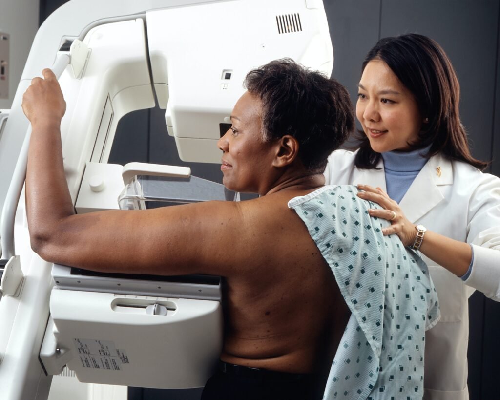
Screening Mammography involves the use of an x-ray using a low-dose of radiation to create an image of the breast. Screening mammograms are used routinely to detect breast cancer in individuals without obvious signs or symptoms of cancer.
Diagnostic Mammography involves an x-ray using a low-dose of radiation to create an image of the breast. It is used to examine breast tissues closely following the appearance of signs of cancer or a suspicious result in a screening mammogram. It can be used to pinpoint an abnormal area to be biopsied (surgically remove tissue) for further examination. It takes longer than a screening mammogram because there are more images of the breast, taken from different angles.
Breast Ultrasounds use high-frequency sound waves to develop images of the breast. It is commonly used in conjunction with mammogram to further examine breast lumps and other abnormalities, to determine if a lump is a tumour or a cyst and to find an abnormal area for a biopsy.
Magnetic Resonance Imaging (MRI) uses magnetic fields to create a three-dimensional image of the breast. MRI’s are not regularly used to diagnose breast cancer, however, they are often used to investigate breast cancer tissues when results from other imaging tests are unclear. They are also often combined with mammography to help detect hereditary breast cancers.
Biopsy – In a biopsy, cells or tissue from a suspicious area of the breast are removed and studied under a microscope to determine if cancer is present. There are many different biopsy methods, but the two most commonly used methods for breast cancer are:
- Core-needle biopsy: A health care provider removes tissues or cells with a needle.
- Surgical biopsy: A surgeon makes a cut (incision) in the breast to remove tissue.
A note about DENSE BREAST
Breast tissue is composed of
- Mix of glandular tissue (responsible for milk production)
- Fibrous tissue (which provides structural support)
- Fatty tissue.
-
Glandular and fibrous tissue vs fatty tissue.
Having dense breasts increases your risk of getting breast cancer as it makes it harder to see a tumour on a mammogram.
Ask your Doctor or Mammogram Technician if you have dense breast – if you do, ask for an ultrasound with your mammogram if you have any concerns or suspicions .

My Personal Experience
I had my Screening Mammography back in September 2021 – the results came back all clear .
When I found my lump in May 2022, my doctor sent me for a Diagnostic Mammogram and Breast Ultrasound.
The results from the two imaging tests lead to a request for me to go for a Biopsy. I was told that from what they could see there was a high probability that I had a tumor – This was the first time I really began to get scared !
I has a Core- Needle Biopsy at the beginning of June 2022 and got my biopsy results back one week later – I had Invasive Ductal Carcinoma – My Cancer Journey begins !!!!
At the time i got my diagnosis i was going through the process of ending my 33 years marriage. I was in complete shock that my Cancer Diagnosis was happening at the same time. I was going to have to walk two major life challenges simultaneously – I kept asking myself – Why Me !! Why Now!!!
Join me as I walk you through the High and Lows of this crazy time .

